Breast cancer remains one of the most pressing global health challenges, with early detection being pivotal to improving survival rates. Despite advances in screening technologies, limitations in sensitivity and the inherent challenges of interpreting imaging mean that some cancers go undetected until they progress further.
A recent study by Hirsch et al. sheds light on the potential of artificial intelligence (AI) to address these gaps, offering new hope for early detection and intervention.
The Motivation Behind the Study
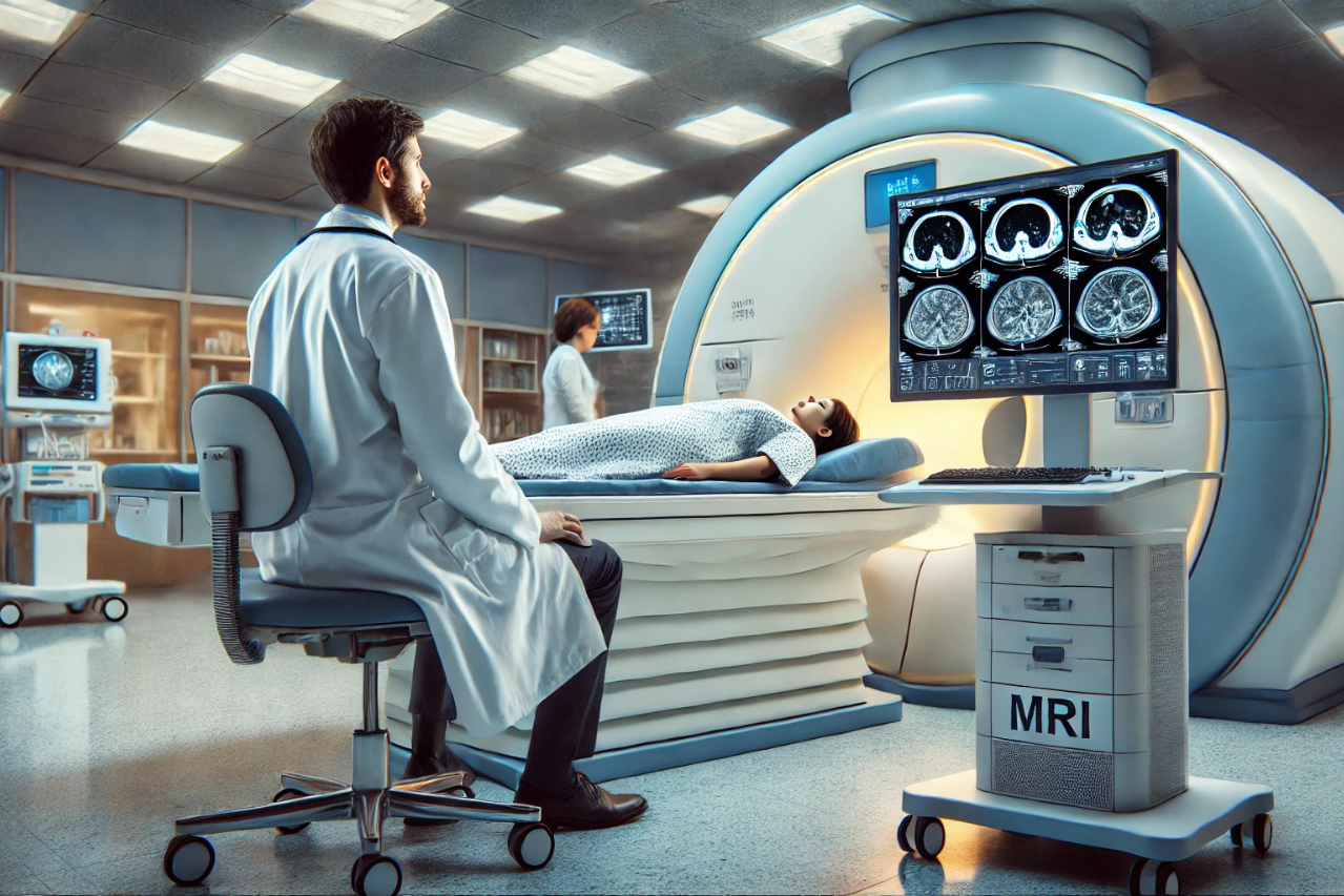
Every year, over 500,000 women in the United States undergo breast magnetic resonance imaging (MRI) as part of supplemental screening programs. These MRIs are particularly valuable for women at higher risk of breast cancer, detecting additional cases missed by traditional mammography. However, even with MRI’s superior sensitivity, a significant percentage of cancers (up to 47% in some studies) may already be present in earlier scans but remain undetected.
The team behind this study hypothesized that AI could analyze these subtle indicators of future cancer development in MRI scans deemed benign by radiologists. The ultimate goal? To predict breast cancer up to a year in advance, giving healthcare professionals a critical head start in treatment planning.
How the Study Was Conducted
Building the AI Model
The researchers employed a convolutional neural network (CNN), a type of AI designed to analyze visual data. This model was initially trained on a vast dataset of breast MRI scans and later fine-tuned using 3,029 MRI images from 910 women, collected over a 12-year period. Notably, the dataset included 115 confirmed cases of breast cancer that developed within a year of initial “negative” screenings.
To overcome challenges such as the low prevalence of cancer in the dataset, the researchers employed advanced techniques like focal loss, which helps the AI focus on identifying rare and subtle indicators of malignancy.
Patient Demographics
This study utilized retrospective data from 910 women who underwent breast MRI screening at a U.S. tertiary cancer center between 2002 and 2014. Participants had consecutive screening exams over an average follow-up period of 4.3 years. The dataset comprised 3,029 sagittal MRI scans, with 115 cancers identified during follow-up.
The study’s participants had a mean age of 52, with MRI scans conducted annually for an average of 4.3 years. Inclusion criteria ensured that the dataset reflected real-world conditions: only scans with BI-RADS scores of 1–3 (indicating benign or probably benign findings) and follow-ups within 15 months. Scans from patients with prior lumpectomies, axillary recurrences, or post-biopsy changes were excluded, resulting in 115 confirmed malignant cases for analysis.
Key Findings
- Improved Early Detection The AI model achieved an area under the receiver operating characteristic curve (AUC-ROC) score of 0.72 for individual breasts, demonstrating its ability to predict cancer development one year in advance. By flagging the 10% of cases deemed most at risk, the model could potentially allow radiologists to detect up to 30% of cancers earlier than traditional methods.
- Pinpointing Cancer Locations One of the most compelling aspects of the study was the AI’s ability to localize future cancer sites. In 57% of cases, the AI correctly identified the anatomical region where cancer would later develop. For true-positive cases flagged for re-evaluation, the success rate rose to an impressive 71%.
- Smaller Lesions, Bigger Impact Many of the cancers detected early were less than 0.5 cm in size—significantly smaller than those typically identified at diagnosis. This highlights the AI’s potential to spot changes that are imperceptible to the human eye but critical for early intervention.
Understanding the Technical Concepts
What Is a Convolutional Neural Network (CNN)?
Imagine flipping through a photo album, looking for changes in a series of similar pictures. A CNN works in much the same way, but with far greater precision. It examines MRI images slice by slice, spotting patterns or abnormalities that could indicate a problem. While radiologists rely on their training and experience, the CNN brings the added advantage of processing immense amounts of data in seconds.
AUC-ROC: The AI Report Card
Think of the AUC-ROC as a scorecard for the AI model. A perfect score of 1.0 means the AI never misses a cancer diagnosis and doesn’t flag any false positives. The model in this study scored 0.72, reflecting a significant improvement over random guessing (0.5) and demonstrating its value as an adjunct to radiologist review.
Balancing Benefits and Costs
The study emphasized the importance of balancing the benefits of early cancer detection with the practical costs of increased radiologist workload. For example:
- A sensitivity of 30% means the AI detects one-third of cancers a year earlier than conventional methods.
- At this sensitivity level, radiologists would need to re-evaluate 10% of cases flagged by AI, translating to a manageable workload increase.
The trade-off is clear: while false positives may slightly increase, the potential to save lives through earlier detection far outweighs this drawback.
Limitations and Future Directions
The study acknowledged several limitations:
- The dataset, while robust, was limited to sagittal MRIs from a single institution.
- Exam-level predictions were less accurate than individual breast-level predictions, highlighting the need for refinement.
Future research will benefit from larger, multi-institutional datasets and higher-resolution imaging technologies. Combining AI predictions with other data, such as genetic risk factors, could further enhance accuracy.
The Broader Impact of AI in Healthcare
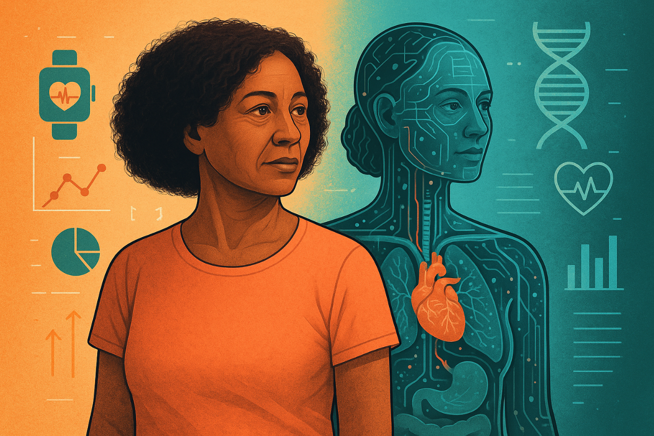
This study is just one example of how AI is transforming medicine. From improving diagnostic accuracy to streamlining workflows, AI has the potential to revolutionize patient care. In breast cancer screening, where early detection can mean the difference between life and death, AI tools like the one developed in this study could set a new standard for precision and reliability.
AI in breast cancer detection represents more than just an incremental improvement—it’s a paradigm shift. By empowering radiologists with advanced tools, we move closer to a future where fewer cancers go undetected, and more lives are saved. As technology continues to evolve, the integration of AI into routine clinical practice promises to make healthcare smarter, more accessible, and, most importantly, more effective.
Are you interested in how AI is changing healthcare? Subscribe to our newsletter, “PulsePoint,” for updates, insights, and trends on AI innovations in healthcare.


